Cardiology prepared for the fourth dimension
DIASTOLE prepares heart surgery by computer simulations in 4D
What if heart specialists could simulate the fitting of a new heart valve in 4D before an operation? 4D CT scanners add the dimension of time to three-dimensional images and visualise the movement of the heart in detail. The imec.icon project DIASTOLE, involving VUB, UZ Brussel and imec, is paving the way to safely implement 4D scans in heart surgery.
The heart is constantly in motion and therefore hard to take images of. During heart surgery it is often difficult to estimate how the heart will react to a new valve. 4D scanners make it possible to check in advance, and thus safely, how a valve will function in a specific patient. 4D imaging is all the more important as today more and more operations are performed via the groin or via a small incision (keyhole technique), whereby the heart is only partly or not directly visible during the operation. To be able to safely use these 4D scanners in cardiac surgery, the various imaging phases had to be revised. The fourth dimension must be added at every stage.
In the imec.icon project DIASTOLE, seven partners worked on those different stages. Researchers from the radiology department of VUB-UZ Brussel developed a model to calculate the radiation dose of 4D scans on the skin, and immediately applied it to draw up a safe protocol. Prof Nico Buls: “For a usable 4D scan, on the one hand the quality has to be sufficient, on the other hand you want to avoid the radiation dose being too high at certain places on the body. Unlike classic CT scans, a 4D scan repeatedly irradiates the same region of the body, so we need to specifically monitor the dose to the skin.”
Prof Jef Vandemeulebroucke, from ETRO, an imec research group at VUB, investigated the image registration and segmentation: “Computers can recognise the heart on a 3D scan and, as it were, separate it from the rest of the body. This way, you get a clear 3D model of the heart. In 4D, you basically have to repeat this for every 3D frame in time. Our approach was to estimate the motion between the different images, compensate for this motion such that you can align and superimpose the different images. Using such a multi-atlas technique, we were able to create high-quality models that our partners in the consortium were able to use in their research.”
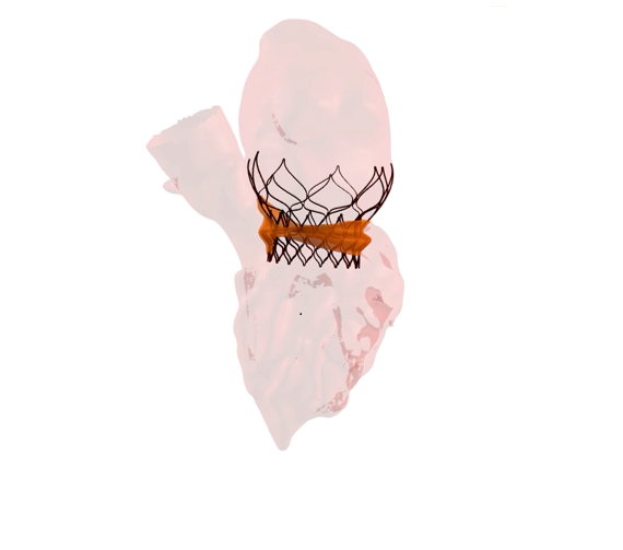
The Centre for Cardiovascular Diseases at UZ Brussel looked at complementarity with 4D ultrasound. GE Healthcare extended its software for radiation dose registration to 4D scans; Materialise did the same for image processing software; Vision Lab, an imec research group from UAntwerp, included 4D in its statistical cardiovascular models. Finally, FEops succeeded in making 4D simulations of a beating heart in which a cardiovascular device be placed virtually. With the application, the doctor can upload 4D images of a patient to a website and have them analysed by FEops. Afterwards, they receive a simulation of the type of implant that would be most appropriate, based on the scan.
“Based on the combined results, I hope we are one step closer to the real use of 4D in cardiac surgery. We now know it is safe, we can process the images and we can make simulations. The approach is feasible, and can be used in clinical applications,” says Vandemeulebroucke.
Future
The 4D scan can also be used for non-patient-specific applications. Provided there is sufficient data, it is also possible to create a representative model with which implants in various sizes can be developed.
Contact
Jef Vandemeulebroucke - General Research Leader
0473 52 46 82
Nico Buls - Research leader radiology VUB-UZ Brussels
0486 512355
Steven Droogmans - Centre for Heart and Vascular Diseases UZ Brussel
0486 95 78 99
Peter Mortier - FEops
0474 27 45 43
DIASTOLE is a collaborative imec.icon research project financed by imec and Agentschap Innoveren & Ondernemen. More information here.
Project partners:
FEops, GE Healthcare DoseWatch Business, Materialise
Research partners:
Centre for Heart and Vascular